|
|
 1. Base Numbering
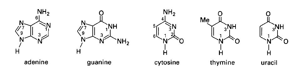 2. Backbone Torsion Angles in Nucleic Acid Structures
atomic numbering scheme and definition of
torsion angles for a polyribonucleotide chain
see also: other information on backbone torsional angles
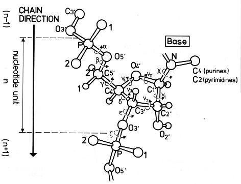
Source: Saenger,W., Principles of Nucleic Acid Structure, Springer
Verlag New York 1984.
| Average Torsion Angles for Nucleic Acid Helices (in °) | |||||||
|---|---|---|---|---|---|---|---|
| Structure Type | Alpha | Beta | Gamma | Delta | Epsilon | Zeta | Chi |
| A-DNA (fibres) | -50 | 172 | 41 | 79 | -146 | -78 | -154 |
| GGCCGGCC | -75 | 185 | 56 | 91 | -166 | -75 | -149 |
| B-DNA (fibres) | -41 | 136 | 38 | 139 | -133 | -157 | -102 |
| CGCGAATTCGCG | -63 | 171 | 54 | 123 | -169 | -108 | -117 |
| Z-DNA (C residues) | -137 | -139 | 56 | 138 | -95 | 80 | -159 |
| Z-DNA (G residues) | 47 | 179 | -169 | 99 | -104 | -69 | 68 |
| DNA-RNA decamer | -69 | 175 | 55 | 82 | -151 | -75 | -162 |
| A-RNA | -68 | 178 | 54 | 82 | -153 | -71 | -158 |
3. Pucker
pseudorotation
cycle of the furanose ring in nucleosides.
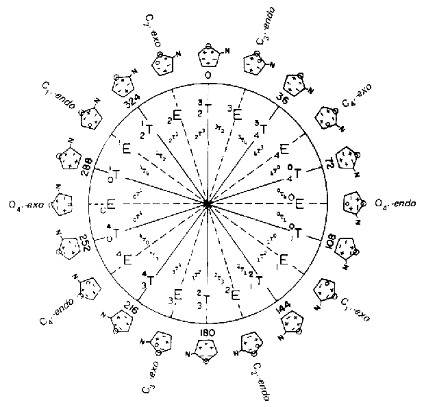
Source: Saenger,W., Principles of
Nucleic Acid Structure, Springer Verlag New York 1984.
ideal B-DNA is C2'-endo (South), ideal A-RNA is C3'-endo (North) 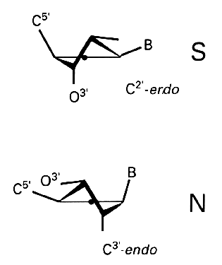
4. Chi Angle
anti and syn conformational ranges
for glycosydic bonds in pyrimidine (left) and purine (right) nucleosides
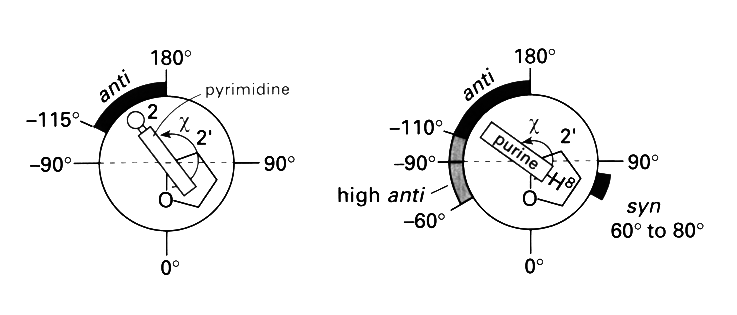
Source: Blackburn and Gait,
Nucleic acids in chemistry and biology, Oxford University Press New
York 1996.
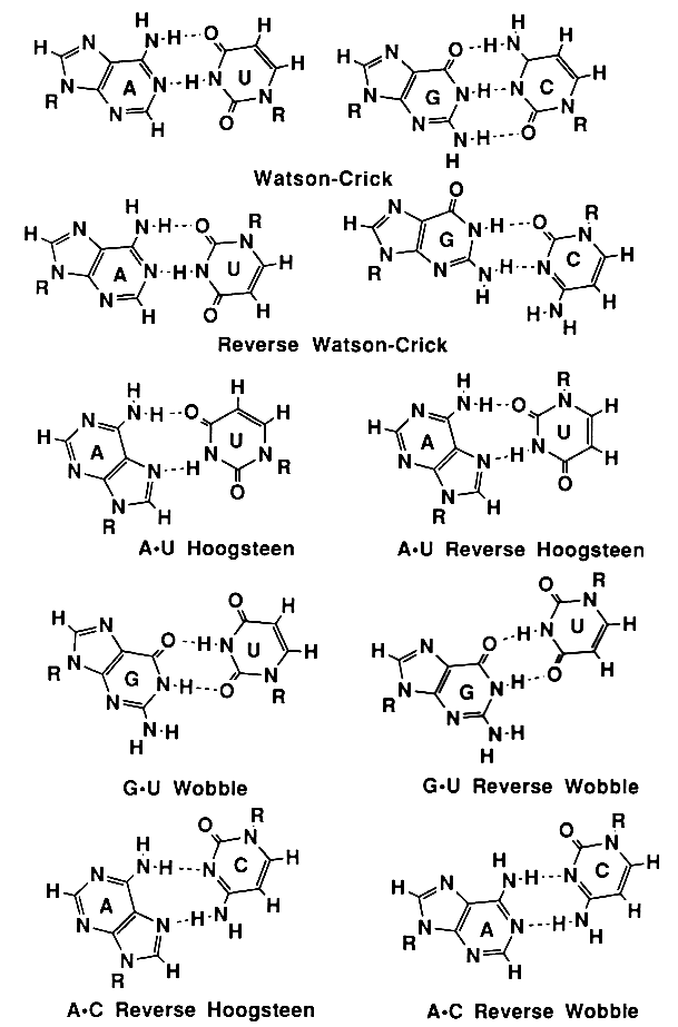
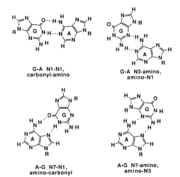
5. Structures of Base Pairs Involving at Least Two Hydrogen Bonds
Watson-Crick, Reverse Watson-Crick, Hoogsteen, Reverse Hoogsteen, Wobble, Reverse Wobble
Homo Purines
the seven
possible homo purine-purine base pairs
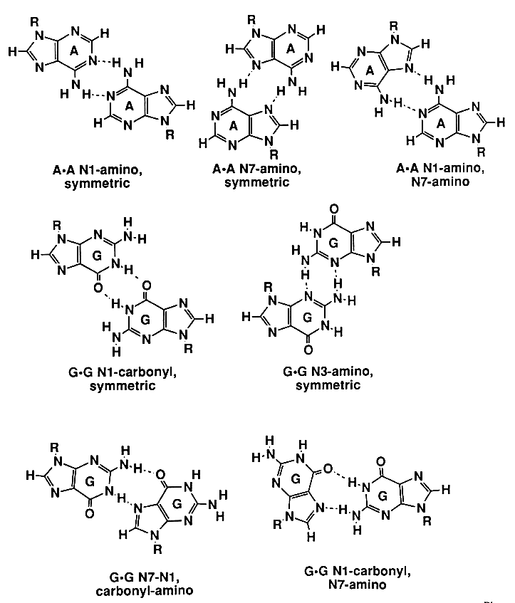
Hetero Purines
the four possible
hetero purine-purine base pairs
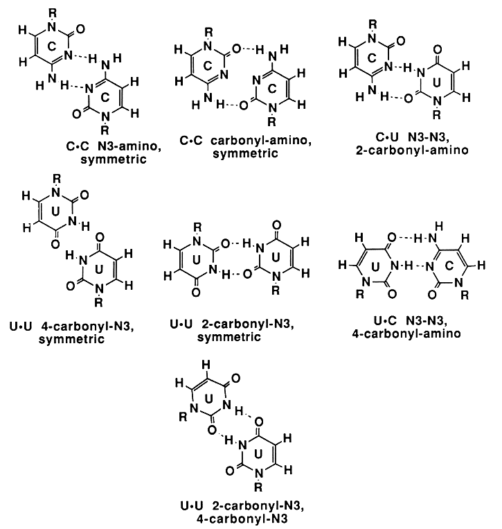
Homo- and Hetero Pyrimidines
the seven possible
pyrimidine-pyrimidine base pairs
6. Base Triples
Proposed
hydrogen-bonding schemes for base triples.
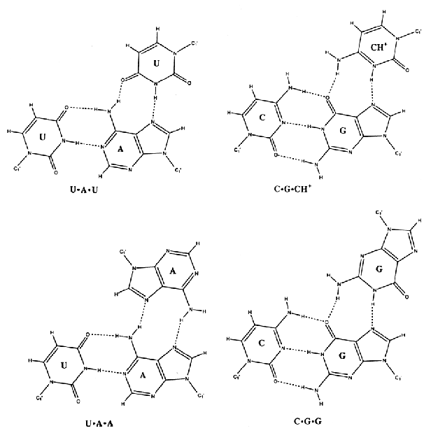
Source: Chastain, M. and Tinoco
Jr., I., (1991) Prog. Nucleic Acid Res. Mol. Biol. 41,
131-177.
7. Movement of Bases(see also: more details on B-DNA and A-RNA)
movements of
bases in sequence-dependent structures (tip, inclination, opening,
propeller, buckle, twist, roll, tilt, slide, rise, shift and tilt).
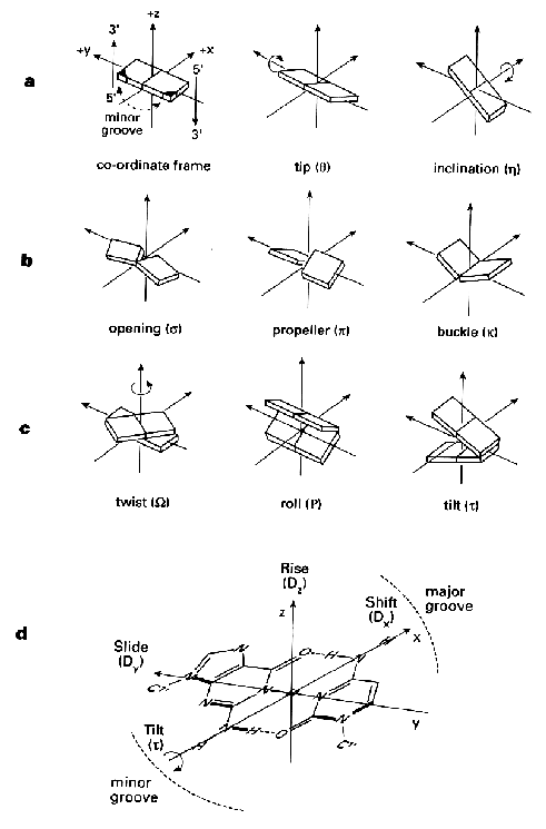
Source: Blackburn and Gait,
Nucleic acids in chemistry and biology, Oxford University Press New
York 1996.
R.E. Dickerson et al. (1989) Nucleic
Acids Res. 17, 1797-1803.
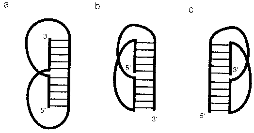
8. Secondary Structures of RNA
secondary
structure of RNA consists of duplex and loop regions that can be
divided into six different types: duplexes, single-stranded regions,
hairpins, internal loops or bubbles, bulge loops or bulges and
junctions.
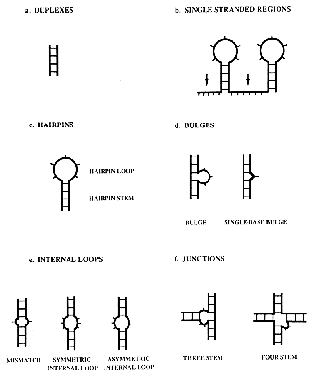
Source: Chastain, M. and Tinoco
Jr., I., (1991) Prog. Nucleic Acid Res. Mol. Biol. 41,
131-177.
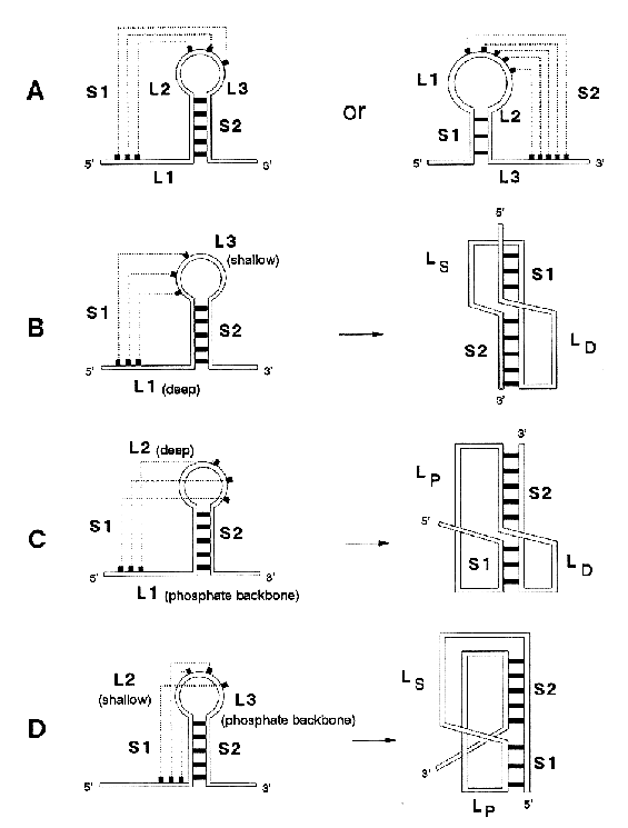
9. PseudoknotsRNA pseudoknots are tertiary structural elements that result when a loop in a secondary structure pairs with a complementary sequence outside the loop
Source: Chastain, M. and Tinoco
Jr., I., (1991) Prog. Nucleic Acid Res. Mol. Biol. 41,
131-177.
The H-type pseudoknot. A pseudoknot is always defined by two stems (S1 and S2) and by two or three loop regions (L1-L3). Dashed lines indicate base-pairing. LD is the loop crossing the deep groove, LS is the loop crossing the shallow groove, and LP is the loop spanning the sugar-phosphate backbone
Source: Cornelis W. A. Pleij in
Gesteland, R. F. and Atkins, J. F. (1993) THE RNA WORLD. Cold Spring
Harbor Laboratory Press.
|
|