|
|
|
|
 Description
|
|
Compounds
|
||||||||||||||||||||||||
Chains, Units
Summary Information (see also Sequences/Alignments below) |
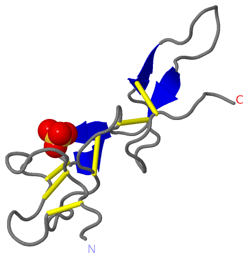
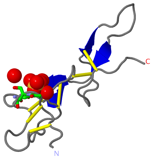
Ligands, Modified Residues, Ions (1, 1)
Asymmetric/Biological Unit (1, 1)
|
Sites (1, 1)
Asymmetric Unit (1, 1)
|
SS Bonds (6, 6)
Asymmetric/Biological Unit
|
||||||||||||||||||||||||||||
Cis Peptide Bonds (0, 0)| (no "Cis Peptide Bond" information available for 1J2L) |
SAPs(SNPs)/Variants (0, 0)| (no "SAP(SNP)/Variant" information available for 1J2L) |
PROSITE Motifs (2, 2)
Asymmetric/Biological Unit (2, 2)
|
||||||||||||||||||||||||||||||||
Exons (0, 0)| (no "Exon" information available for 1J2L) |
Sequences/Alignments
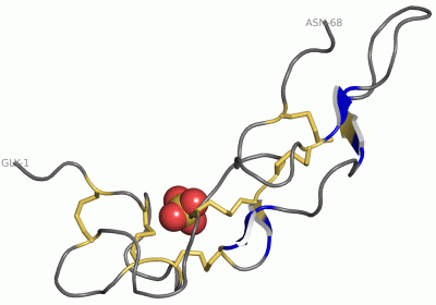
Asymmetric/Biological UnitChain A from PDB Type:PROTEIN Length:68 aligned with VM2T_PROFL | P21859 from UniProtKB/Swiss-Prot Length:70 Alignment length:68 10 20 30 40 50 60 VM2T_PROFL 1 GEECDCGSPSNPCCDAATCKLRPGAQCADGLCCDQCRFKKKRTICRIARGDFPDDRCTGQSADCPRWN 68 SCOP domains d1j2la_ A: Flavoridin (triflavin) SCOP domains CATH domains 1j2lA00 A:1-68 Echistatin CATH domains Pfam domains -------------------------------------------------------------------- Pfam domains SAPs(SNPs) -------------------------------------------------------------------- SAPs(SNPs) PROSITE (1) DISINTEGRIN_2 PDB: A:1-68 UniProt: 1-70 PROSITE (1) PROSITE (2) --------------------------DISINTEGRIN_1 ---------------------- PROSITE (2) Transcript -------------------------------------------------------------------- Transcript 1j2l A 1 GEECDCGSPSNPCCDAATCKLRPGAQCADGLCCDQCRFKKKRTICRIARGDFPDDRCTGQSADCPRWN 68 10 20 30 40 50 60
|
||||||||||||||||||||
SCOP Domains (1, 1)
Asymmetric/Biological Unit
 |
CATH Domains (1, 1)
Asymmetric/Biological Unit
|
Pfam Domains (0, 0)| (no "Pfam Domain" information available for 1J2L) |
Gene Ontology (1, 1)|
Asymmetric/Biological Unit(hide GO term definitions) Chain A (VM2T_PROFL | P21859)
|
||||||||||||
Interactive Views
|
||||||||||||||||||||||||||||||||||||||||||||||||||||||||||||||||||||||||||||||||||||||||||||||||||||||||||||||||||||||
Still Images
|
||||||||||||||||
Databases
|
||||||||||||||||||||||||||||||||||||||||||||||||||||||||||||||||||||||||||||||||||||||||||||||||||||||||||||||||||||||||||||||||||||||||||||||||||||||||||||||||
Analysis Tools
|
|||||||||||||||||||||||||||||||||||||||||||||||||||||||||||||
Entries Sharing at Least One Protein Chain (UniProt ID)
Related Entries Specified in the PDB File
|
|