Show PDB file:   
         Plain Text   HTML   (compressed file size)
QuickSearch:   
by PDB,NDB,UniProt,PROSITE Code or Search Term(s)  
(-)NMR Structure - manually
(-)NMR Structure - model 1
(-)NMR Structure - all models
collapse expand < >
Image NMR Structure - manually
NMR Structure - manually  (Jmol Viewer)
Image NMR Structure - model 1
NMR Structure - model 1  (Jmol Viewer)
Image NMR Structure - all models
NMR Structure - all models  (Jmol Viewer)

(-) Description

Title :  NMR SOLUTION STRUCTURE OF A DISULFIDE ANALOGUE OF THE CYCLIC SUNFLOWER TRYPSIN INHIBITOR SFTI-1
 
Authors :  M. L. J. Korsinczky, R. J. Clark, D. J. Craik
Date :  16 May 04  (Deposition) - 03 May 05  (Release) - 13 Jul 11  (Revision)
Method :  SOLUTION NMR
Resolution :  NOT APPLICABLE
Chains :  NMR Structure  :  A  (20x)
NMR Structure *:  A  (1x)
Keywords :  Sunflower Trypsin Inhibitor, Disulfide Mutant, Hydrolase-Hydrolase Inhibitor Complex (Keyword Search: [Gene Ontology, PubMed, Web (Google))
 
Reference :  M. L. J. Korsinczky, R. J. Clark, D. J. Craik
Disulfide Bond Mutagenesis And The Structure And Function O The Head-To-Tail Macrocyclic Trypsin Inhibitor Sfti-1
Biochemistry V. 44 1145 2005
PubMed-ID: 15667208  |  Reference-DOI: 10.1021/BI048297R

(-) Compounds

Molecule 1 - TRYPSIN INHIBITOR 1
    ChainsA
    EngineeredYES
    MutationYES
    Organism CommonCOMMON SUNFLOWER
    Organism ScientificHELIANTHUS ANNUUS
    Organism Taxid4232
    Other DetailsTHE PEPTIDE WAS SYNTHESIZED USING STANDARD SOLID PHASE PEPTIDE SYNTHESIS METHODS USING BOC CHEMISTRY. THE PEPTIDE BACKBONE WAS CYCLICIZED IN SOLUTION BY ADDITION OF HBTU AND DIEA WHICH RESULTED IN THE FORMATION OF A PEPTIDE BOND BETWEEN D14 AND G1. THE SEQUENCE OF THE PEPTIDE IS NATURALLY FOUND IN HELIANTHUS ANNUS (SUNFLOWER)
    SynonymSFTI-1
    SyntheticYES

 Structural Features

(-) Chains, Units

  1
NMR Structure (20x)A
NMR Structure * (1x)A

Summary Information (see also Sequences/Alignments below)

(-) Ligands, Modified Residues, Ions  (1, 2)

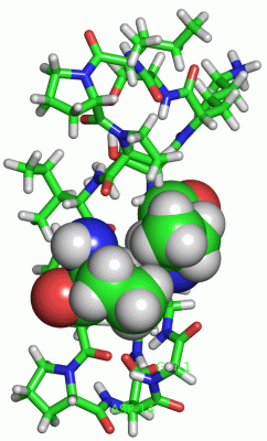
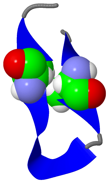
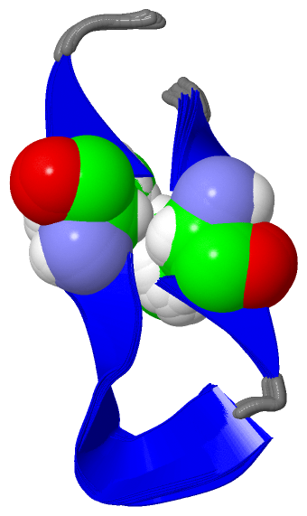
NMR Structure (1, 2)
No.NameCountTypeFull Name
1ABA2Mod. Amino AcidALPHA-AMINOBUTYRIC ACID
NMR Structure * (1, 2)
No.NameCountTypeFull Name
1ABA2Mod. Amino AcidALPHA-AMINOBUTYRIC ACID

(-) Sites  (0, 0)

(no "Site" information available for 1T9E)

(-) SS Bonds  (0, 0)

(no "SS Bond" information available for 1T9E)

(-) Cis Peptide Bonds  (1, 20)

NMR Structure
No.ModelResidues
11, 2, 3, 4, 5, 6, 7, 8, 9, 10, 11, 12, 13, 14, 15, 16, 17, 18, 19, 20Ile A:7 -Pro A:8

 Sequence-Structure Mapping

(-) SAPs(SNPs)/Variants  (0, 0)

(no "SAP(SNP)/Variant" information available for 1T9E)

(-) PROSITE Motifs  (0, 0)

(no "PROSITE Motif" information available for 1T9E)

(-) Exons   (0, 0)

(no "Exon" information available for 1T9E)

(-) Sequences/Alignments

NMR Structure
   Reformat: Number of residues per line =  ('0' or empty: single-line sequence representation)
  Number of residues per labelling interval =   
  UniProt sequence: complete  aligned part    
   Show mapping: SCOP domains CATH domains Pfam domains Secondary structure (by author)
SAPs(SNPs) PROSITE motifs Exons
(details for a mapped element are shown in a popup box when the mouse pointer rests over it)
Chain A from PDB  Type:PROTEIN  Length:14
 aligned with SFTI1_HELAN | Q4GWU5 from UniProtKB/Swiss-Prot  Length:56

    Alignment length:14
                                    49    
           SFTI1_HELAN   40 GRCTKSIPPICFPD 53
               SCOP domains -------------- SCOP domains
               CATH domains -------------- CATH domains
               Pfam domains -------------- Pfam domains
         Sec.struct. author .............. Sec.struct. author
                 SAPs(SNPs) -------------- SAPs(SNPs)
                    PROSITE -------------- PROSITE
                 Transcript -------------- Transcript
                  1t9e A  1 GRaTKSIPPIaFPD 14
                              |     10|   
                              |      11-ABA
                              3-ABA       

   Legend:   → Mismatch (orange background)
  - → Gap (green background, '-', border residues have a numbering label)
    → Modified Residue (blue background, lower-case, 'x' indicates undefined single-letter code, labelled with number + name)
  x → Chemical Group (purple background, 'x', labelled with number + name, e.g. ACE or NH2)
  extra numbering lines below/above indicate numbering irregularities and modified residue names etc., number ends below/above '|'

 Classification and Annotation

(-) SCOP Domains  (0, 0)

(no "SCOP Domain" information available for 1T9E)

(-) CATH Domains  (0, 0)

(no "CATH Domain" information available for 1T9E)

(-) Pfam Domains  (0, 0)

(no "Pfam Domain" information available for 1T9E)

(-) Gene Ontology  (4, 4)

NMR Structure(hide GO term definitions)
Chain A   (SFTI1_HELAN | Q4GWU5)
molecular function
    GO:0030414    peptidase inhibitor activity    Stops, prevents or reduces the activity of a peptidase, any enzyme that catalyzes the hydrolysis peptide bonds.
    GO:0004867    serine-type endopeptidase inhibitor activity    Stops, prevents or reduces the activity of serine-type endopeptidases, enzymes that catalyze the hydrolysis of nonterminal peptide bonds in a polypeptide chain; a serine residue (and a histidine residue) are at the active center of the enzyme.
biological process
    GO:0010951    negative regulation of endopeptidase activity    Any process that decreases the frequency, rate or extent of endopeptidase activity, the endohydrolysis of peptide bonds within proteins.
    GO:0010466    negative regulation of peptidase activity    Any process that stops or reduces the rate of peptidase activity, the hydrolysis of peptide bonds within proteins.

 Visualization

(-) Interactive Views

NMR Structure
  Complete Structure
    Jena3D(integrated viewing of ligand, site, SAP, PROSITE, SCOP information)
    WebMol | AstexViewer[tm]@PDBe
(Java Applets, require no local installation except for Java; loading may be slow)
    STRAP
(Java WebStart application, automatic local installation, requires Java; full application with system access!)
    RasMol
(require local installation)
    Molscript (VRML)
(requires installation of a VRML viewer; select preferred view via VRML and generate a mono or stereo PDF format file)
 
  Ligands, Modified Residues, Ions
    ABA  [ RasMol | Jena3D ]  +environment [ RasMol | Jena3D ]
 
  Sites
(no "Sites" information available for 1t9e)
 
  Cis Peptide Bonds
    Ile A:7 - Pro A:8   [ RasMol ]  
 

(-) Still Images

Jmol
  protein: cartoon or spacefill or dots and stick; nucleic acid: cartoon and stick; ligands: spacefill; active site: stick
Molscript
  protein, nucleic acid: cartoon; ligands: spacefill; active site: ball and stick

 Databases and Analysis Tools

(-) Databases

Access by PDB/NDB ID
  1t9e
    Family and Domain InformationProDom | SYSTERS
    General Structural InformationGlycoscienceDB | MMDB | NDB | OCA | PDB | PDBe | PDBj | PDBsum | PDBWiki | PQS | PROTEOPEDIA
    Orientation in MembranesOPM
    Protein SurfaceSURFACE
    Secondary StructureDSSP (structure derived) | HSSP (homology derived)
    Structural GenomicsGeneCensus
    Structural NeighboursCE | VAST
    Structure ClassificationCATH | Dali | SCOP
    Validation and Original DataBMRB Data View | BMRB Restraints Grid | EDS | PROCHECK | RECOORD | WHAT_CHECK
 
Access by UniProt ID/Accession number
  SFTI1_HELAN | Q4GWU5
    Comparative Protein Structure ModelsModBase
    Genomic InformationEnsembl
    Protein-protein InteractionDIP
    Sequence, Family and Domain InformationInterPro | Pfam | SMART | UniProtKB/SwissProt
 
Access by Enzyme Classificator   (EC Number)
  (no 'Enzyme Classificator' available)
    General Enzyme InformationBRENDA | EC-PDB | Enzyme | IntEnz
    PathwayKEGG | MetaCyc
 
Access by Disease Identifier   (MIM ID)
  (no 'MIM ID' available)
    Disease InformationOMIM
 
Access by GenAge ID
  (no 'GenAge ID' available)
    Age Related InformationGenAge

(-) Analysis Tools

Access by PDB/NDB ID
    Domain InformationXDom
    Interatomic Contacts of Structural UnitsCSU
    Ligand-protein ContactsLPC
    Protein CavitiescastP
    Sequence and Secondary StructurePDBCartoon
    Structure AlignmentSTRAP(Java WebStart application, automatic local installation, requires Java; full application with system access!)
    Structure and Sequence BrowserSTING
 
Access by UniProt ID/Accession number
  SFTI1_HELAN | Q4GWU5
    Protein Disorder PredictionDisEMBL | FoldIndex | GLOBPLOT (for more information see DisProt)

 Related Entries

(-) Entries Sharing at Least One Protein Chain (UniProt ID)

UniProtKB/Swiss-Prot
        SFTI1_HELAN | Q4GWU51jbl 1jbn 1o8y 1o8z 1sfi 2ab9 3p8f 4abi 4abj 4hgc 4k1e 4k8y 4kel 4xoj

(-) Related Entries Specified in the PDB File

1jbl NMR STRUCTURE OF NATIVE SFTI-1
1jbn NMR STRUCTURE OF AN ACYCLIC PERMUTANT OF SFTI-1
1o8y NMR STRUCTURE OF AN ACYCLIC PERMUTANT OF SFTI-1
1o8z NMR STRUCTURE OF AN ACYCLIC PERMUTANT OF SFTI-1
1sfi CRYSTAL STRUCTURE OF NATIVE SFTI-1