Show PDB file:   
         Plain Text   HTML   (compressed file size)
QuickSearch:   
by PDB,NDB,UniProt,PROSITE Code or Search Term(s)  
(-)Asym./Biol. Unit
(-)Asym./Biol. Unit - sites
collapse expand < >
Image Asym./Biol. Unit
Asym./Biol. Unit  (Jmol Viewer)
Image Asym./Biol. Unit - sites
Asym./Biol. Unit - sites  (Jmol Viewer)

(-) Description

Title :  THE PR1 PHOTOCYCLE INTERMEDIATE OF PHOTOACTIVE YELLOW PROTEIN
 
Authors :  F. Schotte, H. S. Cho, V. R. I. Kaila, H. Kamikubo, N. Dashdorj, E. R. Henr T. J. Graber, R. Henning, M. Wulff, G. Hummer, M. Kataoka, P. A. Anfinru
Date :  27 Sep 12  (Deposition) - 14 Nov 12  (Release) - 25 Jan 17  (Revision)
Method :  X-RAY DIFFRACTION
Resolution :  1.60
Chains :  Asym./Biol. Unit :  A
Keywords :  Photoreceptor, Light Sensor, Chromophore, Photoreceptor Protein, Receptor, Sensory Transduction, Signaling Protein, Pas, Lov (Keyword Search: [Gene Ontology, PubMed, Web (Google))
 
Reference :  F. Schotte, H. S. Cho, V. R. I. Kaila, H. Kamikubo, N. Dashdorj, E. R. Henry, T. J. Graber, R. Henning, M. Wulff, G. Hummer, M. Kataoka, P. A. Anfinrud
Watching A Signaling Protein Function In Real Time Via 100-Ps Time-Resolved Laue Crystallography.
Proc. Natl. Acad. Sci. Usa V. 109 19256 2012
PubMed-ID: 23132943  |  Reference-DOI: 10.1073/PNAS.1210938109

(-) Compounds

Molecule 1 - PHOTOACTIVE YELLOW PROTEIN
    ChainsA
    EngineeredYES
    Expression SystemESCHERICHIA COLI
    Expression System PlasmidPET16B
    Expression System StrainBL21(DE3)
    Expression System Taxid469008
    Expression System Vector TypePLASMID
    Organism ScientificHALORHODOSPIRA HALOPHILA
    Organism Taxid1053
    SynonymPYP

 Structural Features

(-) Chains, Units

  1
Asymmetric/Biological Unit A

Summary Information (see also Sequences/Alignments below)

(-) Ligands, Modified Residues, Ions  (1, 1)

Asymmetric/Biological Unit (1, 1)
No.NameCountTypeFull Name
1HC41Ligand/Ion4'-HYDROXYCINNAMIC ACID

(-) Sites  (1, 1)

Asymmetric Unit (1, 1)
No.NameEvidenceResiduesDescription
1AC1SOFTWARETYR A:42 , GLU A:46 , THR A:50 , ARG A:52 , VAL A:66 , ALA A:67 , CYS A:69 , THR A:70 , PHE A:96 , TYR A:98BINDING SITE FOR RESIDUE HC4 A 169

(-) SS Bonds  (0, 0)

(no "SS Bond" information available for 4BBT)

(-) Cis Peptide Bonds  (0, 0)

(no "Cis Peptide Bond" information available for 4BBT)

 Sequence-Structure Mapping

(-) SAPs(SNPs)/Variants  (0, 0)

(no "SAP(SNP)/Variant" information available for 4BBT)

(-) PROSITE Motifs  (1, 1)

Asymmetric/Biological Unit (1, 1)
 PROSITEUniProtKBPDB
No.IDACDescriptionIDLocationCountLocation
1PASPS50112 PAS repeat profile.PYP_HALHA23-86  1A:23-86

(-) Exons   (0, 0)

(no "Exon" information available for 4BBT)

(-) Sequences/Alignments

Asymmetric/Biological Unit
   Reformat: Number of residues per line =  ('0' or empty: single-line sequence representation)
  Number of residues per labelling interval =   
  UniProt sequence: complete  aligned part    
   Show mapping: SCOP domains CATH domains Pfam domains Secondary structure (by author)
SAPs(SNPs) PROSITE motifs Exons
(details for a mapped element are shown in a popup box when the mouse pointer rests over it)
Chain A from PDB  Type:PROTEIN  Length:125
 aligned with PYP_HALHA | P16113 from UniProtKB/Swiss-Prot  Length:125

    Alignment length:125
                                    10        20        30        40        50        60        70        80        90       100       110       120     
            PYP_HALHA     1 MEHVAFGSEDIENTLAKMDDGQLDGLAFGAIQLDGDGNILQYNAAEGDITGRDPKQVIGKNFFKDVAPCTDSPEFYGKFKEGVASGNLNTMFEYTFDYQMTPTKVKVHMKKALSGDSYWVFVKRV 125
               SCOP domains d4bbta_ A: Photoactive yellow protein, PYP                                                                                    SCOP domains
               CATH domains ----------------------------------------------------------------------------------------------------------------------------- CATH domains
               Pfam domains ----------------------------------------------------------------------------------------------------------------------------- Pfam domains
         Sec.struct. author .........hhhhhhh..hhhhhh....eeeeee....eeeehhhhhhhhh.hhhhhh...hhhhhhhhhh...hhhhhhhhhhhh..eeeeeeee......eeeeeeeee.....eeeeeeee. Sec.struct. author
                 SAPs(SNPs) ----------------------------------------------------------------------------------------------------------------------------- SAPs(SNPs)
                    PROSITE ----------------------PAS  PDB: A:23-86 UniProt: 23-86                                --------------------------------------- PROSITE
                 Transcript ----------------------------------------------------------------------------------------------------------------------------- Transcript
                 4bbt A   1 MEHVAFGSEDIENTLAKMDDGQLDGLAFGAIQLDGDGNILQYNAAEGDITGRDPKQVIGKNFFKDVAPCTDSPEFYGKFKEGVASGNLNTMFEYTFDYQMTPTKVKVHMKKALSGDSYWVFVKRV 125
                                    10        20        30        40        50        60        70        80        90       100       110       120     

   Legend:   → Mismatch (orange background)
  - → Gap (green background, '-', border residues have a numbering label)
    → Modified Residue (blue background, lower-case, 'x' indicates undefined single-letter code, labelled with number + name)
  x → Chemical Group (purple background, 'x', labelled with number + name, e.g. ACE or NH2)
  extra numbering lines below/above indicate numbering irregularities and modified residue names etc., number ends below/above '|'

 Classification and Annotation

(-) SCOP Domains  (1, 1)

Asymmetric/Biological Unit

(-) CATH Domains  (0, 0)

(no "CATH Domain" information available for 4BBT)

(-) Pfam Domains  (0, 0)

(no "Pfam Domain" information available for 4BBT)

(-) Gene Ontology  (5, 5)

Asymmetric/Biological Unit(hide GO term definitions)
Chain A   (PYP_HALHA | P16113)
molecular function
    GO:0009881    photoreceptor activity    The function of absorbing and responding to incidental electromagnetic radiation, particularly visible light. The response may involve a change in conformation.
biological process
    GO:0007602    phototransduction    The sequence of reactions within a cell required to convert absorbed photons into a molecular signal.
    GO:0018298    protein-chromophore linkage    The covalent or noncovalent attachment of a chromophore to a protein.
    GO:0006355    regulation of transcription, DNA-templated    Any process that modulates the frequency, rate or extent of cellular DNA-templated transcription.
    GO:0050896    response to stimulus    Any process that results in a change in state or activity of a cell or an organism (in terms of movement, secretion, enzyme production, gene expression, etc.) as a result of a stimulus. The process begins with detection of the stimulus and ends with a change in state or activity or the cell or organism.

 Visualization

(-) Interactive Views

Asymmetric/Biological Unit
  Complete Structure
    Jena3D(integrated viewing of ligand, site, SAP, PROSITE, SCOP information)
    WebMol | AstexViewer[tm]@PDBe
(Java Applets, require no local installation except for Java; loading may be slow)
    STRAP
(Java WebStart application, automatic local installation, requires Java; full application with system access!)
    RasMol
(require local installation)
    Molscript (VRML)
(requires installation of a VRML viewer; select preferred view via VRML and generate a mono or stereo PDF format file)
 
  Ligands, Modified Residues, Ions
    HC4  [ RasMol | Jena3D ]  +environment [ RasMol | Jena3D ]
 
  Sites
    AC1  [ RasMol ]  +environment [ RasMol ]
 
  Cis Peptide Bonds
(no "Cis Peptide Bonds" information available for 4bbt)
 

(-) Still Images

Jmol
  protein: cartoon or spacefill or dots and stick; nucleic acid: cartoon and stick; ligands: spacefill; active site: stick
Molscript
  protein, nucleic acid: cartoon; ligands: spacefill; active site: ball and stick

 Databases and Analysis Tools

(-) Databases

Access by PDB/NDB ID
  4bbt
    Family and Domain InformationProDom | SYSTERS
    General Structural InformationGlycoscienceDB | MMDB | NDB | OCA | PDB | PDBe | PDBj | PDBsum | PDBWiki | PQS | PROTEOPEDIA
    Orientation in MembranesOPM
    Protein SurfaceSURFACE
    Secondary StructureDSSP (structure derived) | HSSP (homology derived)
    Structural GenomicsGeneCensus
    Structural NeighboursCE | VAST
    Structure ClassificationCATH | Dali | SCOP
    Validation and Original DataBMRB Data View | BMRB Restraints Grid | EDS | PROCHECK | RECOORD | WHAT_CHECK
 
Access by UniProt ID/Accession number
  PYP_HALHA | P16113
    Comparative Protein Structure ModelsModBase
    Genomic InformationEnsembl
    Protein-protein InteractionDIP
    Sequence, Family and Domain InformationInterPro | Pfam | SMART | UniProtKB/SwissProt
 
Access by Enzyme Classificator   (EC Number)
  (no 'Enzyme Classificator' available)
    General Enzyme InformationBRENDA | EC-PDB | Enzyme | IntEnz
    PathwayKEGG | MetaCyc
 
Access by Disease Identifier   (MIM ID)
  (no 'MIM ID' available)
    Disease InformationOMIM
 
Access by GenAge ID
  (no 'GenAge ID' available)
    Age Related InformationGenAge

(-) Analysis Tools

Access by PDB/NDB ID
    Domain InformationXDom
    Interatomic Contacts of Structural UnitsCSU
    Ligand-protein ContactsLPC
    Protein CavitiescastP
    Sequence and Secondary StructurePDBCartoon
    Structure AlignmentSTRAP(Java WebStart application, automatic local installation, requires Java; full application with system access!)
    Structure and Sequence BrowserSTING
 
Access by UniProt ID/Accession number
  PYP_HALHA | P16113
    Protein Disorder PredictionDisEMBL | FoldIndex | GLOBPLOT (for more information see DisProt)

 Related Entries

(-) Entries Sharing at Least One Protein Chain (UniProt ID)

UniProtKB/Swiss-Prot
        PYP_HALHA | P161131d7e 1f98 1f9i 1gsv 1gsw 1gsx 1kou 1nwz 1odv 1ot6 1ot9 1ota 1otb 1otd 1ote 1oti 1s1y 1s1z 1s4r 1s4s 1t18 1t19 1t1a 1t1b 1t1c 1ts0 1ts6 1ts7 1ts8 1ugu 1uwn 1uwp 1xfn 1xfq 2d01 2d02 2i9v 2kx6 2phy 2pyp 2pyr 2qj5 2qj7 2qws 2zoh 2zoi 3phy 3pyp 3umd 3ume 3ve3 3ve4 4b9o 4bbu 4bbv 4hy8 4i38 4i39 4i3a 4i3i 4i3j 4wl9 4wla 5hd3 5hd5 5hdc 5hdd 5hds

(-) Related Entries Specified in the PDB File

1d7e CRYSTAL STRUCTURE OF THE P65 CRYSTAL FORM OF PHOTOACTIVEYELLOW PROTEIN
1f98 CRYSTAL STRUCTURE OF THE PHOTOACTIVE YELLOW PROTEIN MUTANTT50V
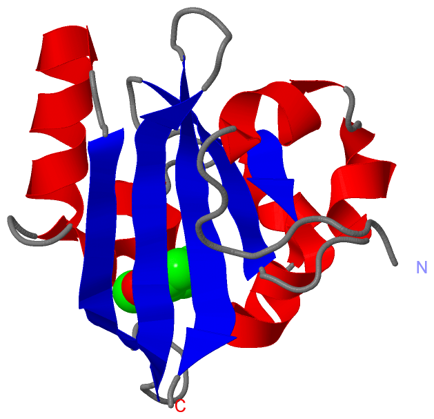
1f9i CRYSTAL STRUCTURE OF THE PHOTOACTIVE YELLOW PROTEIN MUTANTY42F
1gsv CRYSTAL STRUCTURE OF THE P65 CRYSTAL FORM OF PHOTOACTIVE YELLOW PROTEIN
1gsw CRYSTAL STRUCTURE OF THE P65 CRYSTAL FORM OF PHOTOACTIVE YELLOW PROTEIN G51S MUTANT
1gsx CRYSTAL STRUCTURE OF THE P65 CRYSTAL FORM OF PHOTOACTIVE YELLOW PROTEIN G47S/G51S MUTANT
1kou CRYSTAL STRUCTURE OF THE PHOTOACTIVE YELLOW PROTEINRECONSTITUTED WITH CAFFEIC ACID AT 1.16 A RESOLUTION
1nwz PYP ULTRA-HIGH RESOLUTION STRUCTURE OF A BACTERIALPHOTORECEPTOR
1odv PHOTOACTIVE YELLOW PROTEIN 1-25 DELETION MUTANT
1ot6 CRYOTRAPPED CRYSTAL STRUCTURE OF THE E46Q MUTANT OFPHOTOACTIVE YELLOW PROTEIN UNDER CONTINUOUS ILLUMINATIONAT 110K
1ot9 CRYOTRAPPED STATE IN WILD TYPE PHOTOACTIVE YELLOW PROTEIN,INDUCED WITH CONTINUOUS ILLUMINATION AT 110K
1ota E46Q MUTANT OF PHOTOACTIVE YELLOW PROTEIN, P63 AT 295K
1otb WILD TYPE PHOTOACTIVE YELLOW PROTEIN, P63 AT 295K
1otd STRONG HYDROGEN BONDS IN PHOTOACTIVE YELLOW PROTEIN ANDTHEIR ROLE IN ITS PHOTOCYCLE
1ote E46Q MUTANT OF PHOTOACTIVE YELLOW PROTEIN, P65 AT 110K
1oti E46Q MUTANT OF PHOTOACTIVE YELLOW PROTEIN, P65 AT 295K
1s1y PHOTOACTIVATED CHROMOPHORE CONFORMATION IN PHOTOACTIVEYELLOW PROTEIN (E46Q MUTANT) FROM 10 MICROSECONDS TO 3MILLISECONDS
1s1z PHOTOACTIVATED CHROMOPHORE CONFORMATION IN PHOTOACTIVEYELLOW PROTEIN (E46Q MUTANT) FROM 10 TO 500 NANOSECONDS
1s4r STRUCTURE OF A REACTION INTERMEDIATE IN THE PHOTOCYCLE OFPYP EXTRACTED BY A SVD-DRIVEN ANALYSIS
1s4s REACTION INTERMEDIATE IN THE PHOTOCYCLE OF PYP, INTERMEDIATE OCCUPIED BETWEEN 100 MICRO-SECONDS TO 5 MILLI-SECONDS
1t18 EARLY INTERMEDIATE IE1 FROM TIME-RESOLVED CRYSTALLOGRAPHYOF THE E46Q MUTANT OF PYP
1t19 EARLY INTERMEDIATE IE2 FROM TIME-RESOLVED CRYSTALLOGRAPHYOF THE E46Q MUTANT OF PYP
1t1a LATE INTERMEDIATE IL1 FROM TIME-RESOLVED CRYSTALLOGRAPHY OFTHE E46Q MUTANT OF PYP
1t1b LATE INTERMEDIATE IL2 FROM TIME-RESOLVED CRYSTALLOGRAPHY OFTHE E46Q MUTANT OF PYP
1t1c LATE INTERMEDIATE IL3 FROM TIME-RESOLVED CRYSTALLOGRAPHY OFTHE E46Q MUTANT OF PYP
1ts0 STRUCTURE OF THE PB1 INTERMEDIATE FROM TIME-RESOLVED LAUECRYSTALLOGRAPHY
1ts6 STRUCTURE OF THE PB2 INTERMEDIATE FROM TIME-RESOLVED LAUECRYSTALLOGRAPHY
1ts7 STRUCTURE OF THE PR CIS WOBBLE AND PR E46Q INTERMEDIATESFROM TIME- RESOLVED LAUE CRYSTALLOGRAPHY
1ts8 STRUCTURE OF THE PR CIS PLANAR INTERMEDIATE FROM TIME -RESOLVED LAUE CRYSTALLOGRAPHY
1ugu CRYSTAL STRUCTURE OF PYP E46Q MUTANT
1uwn THE INITIAL EVENTS IN THE PHOTOCYCLE OF PHOTOACTIVE YELLOW PROTEIN: A COMMON MECHANISM ON LIGHT ACTIVATION IN PHOTORECEPTOR PROTEINS
1uwp INITIAL EVENTS IN THE PHOTOCYCLE OF PHOTOACTIVE YELLOW PROTEIN
1xfn NMR STRUCTURE OF THE GROUND STATE OF THE PHOTOACTIVE YELLOWPROTEIN LACKING THE N-TERMINAL PART
1xfq STRUCTURE OF THE BLUE SHIFTED INTERMEDIATE STATE OF THEPHOTOACTIVE YELLOW PROTEIN LACKING THE N-TERMINAL PART
2d01 WILD TYPE PHOTOACTIVE YELLOW PROTEIN, P65 FORM
2d02 R52Q MUTANT OF PHOTOACTIVE YELLOW PROTEIN, P65 FORM
2phy PHOTOACTIVE YELLOW PROTEIN, DARK STATE (UNBLEACHED) 2PHY 3
2pyp PHOTOACTIVE YELLOW PROTEIN, PHOTOSTATIONARY STATE, 50% GROUND STATE, 50% BLEACHED
2pyr PHOTOACTIVE YELLOW PROTEIN, 1 NANOSECOND INTERMEDIATE (287K)
3phy PHOTOACTIVE YELLOW PROTEIN, DARK STATE (UNBLEACHED), SOLUTION STRUCTURE, NMR, 26 STRUCTURES
3pyp PHOTOACTIVE YELLOW PROTEIN, CRYOTRAPPED EARLY LIGHT CYCLEINTERMEDIATE
4b9o THE PR0 PHOTOCYCLE INTERMEDIATE OF PHOTOACTIVE YELLOW PROTEIN
4bbu THE PR1 PHOTOCYCLE INTERMEDIATE OF PHOTOACTIVE YELLOW PROTEIN
4bbv THE PB0 PHOTOCYCLE INTERMEDIATE OF PHOTOACTIVE YELLOW PROTEIN