 |
Three-Dimensional Structures of Peptaibols |
Cephaibol B
Sequence:
| Ac |
PHE 01 |
AIB 02 |
AIB 03 |
AIB 04 |
IVA 05 |
GLY 06 |
LEU 07 |
IVA 08 |
AIB 09 |
HYP 10 |
|
GLN 11 |
IVA 12 |
HYP 13 |
AIB 14 |
PRO 15 |
PHE 16 |
ol |
- |
- |
- |
Structural Information (X-ray structure, resolution: 0.89 Å, 2
chains):
 |
-
PDB file (PDB
code: 1OB6)
-
JenaLib atlas page (offers access to PDB, PDBSUM, OCA, MMDB,
... and a variety of visualizations)
-
Bending analysis
-
Methods
-
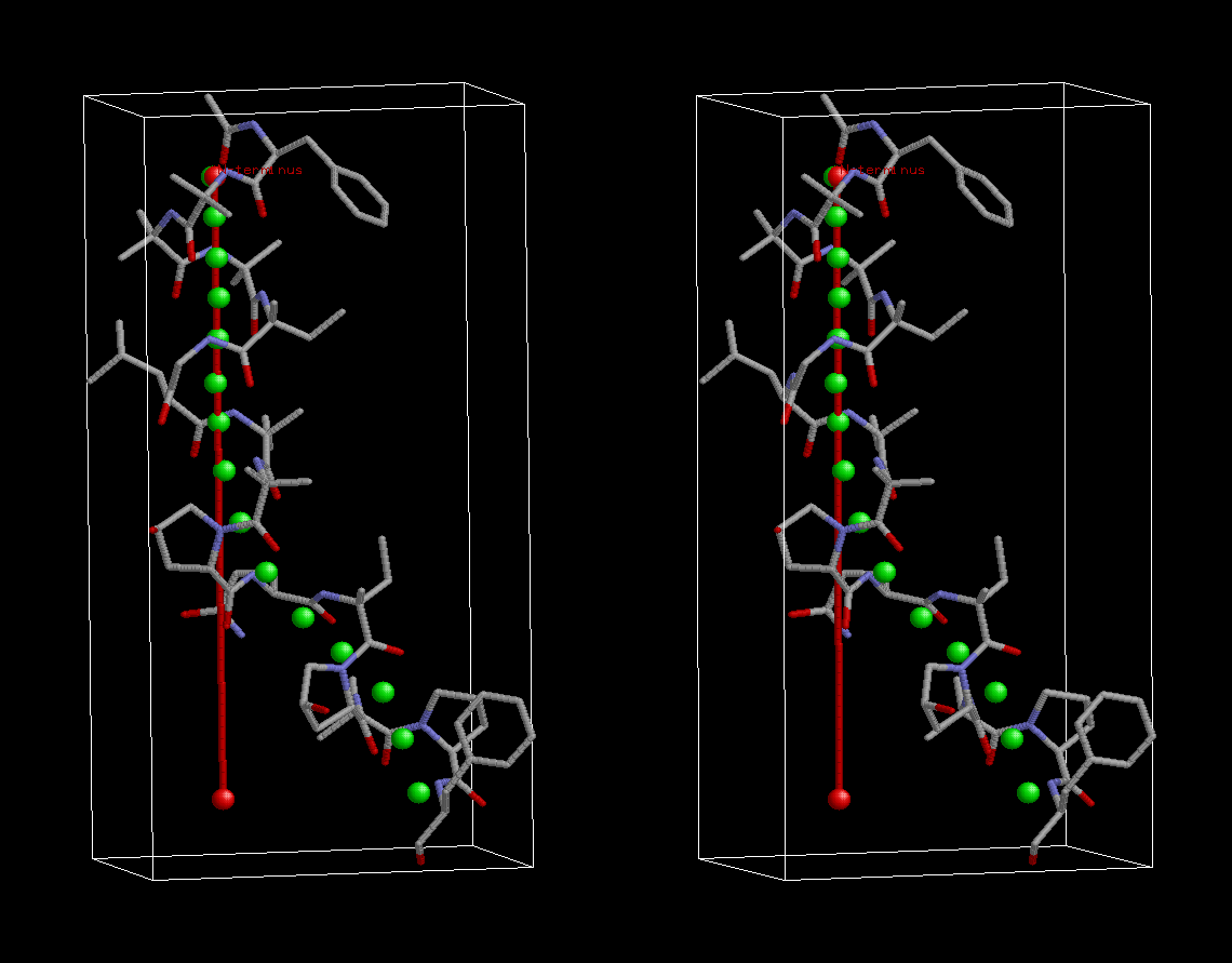
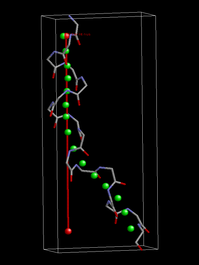
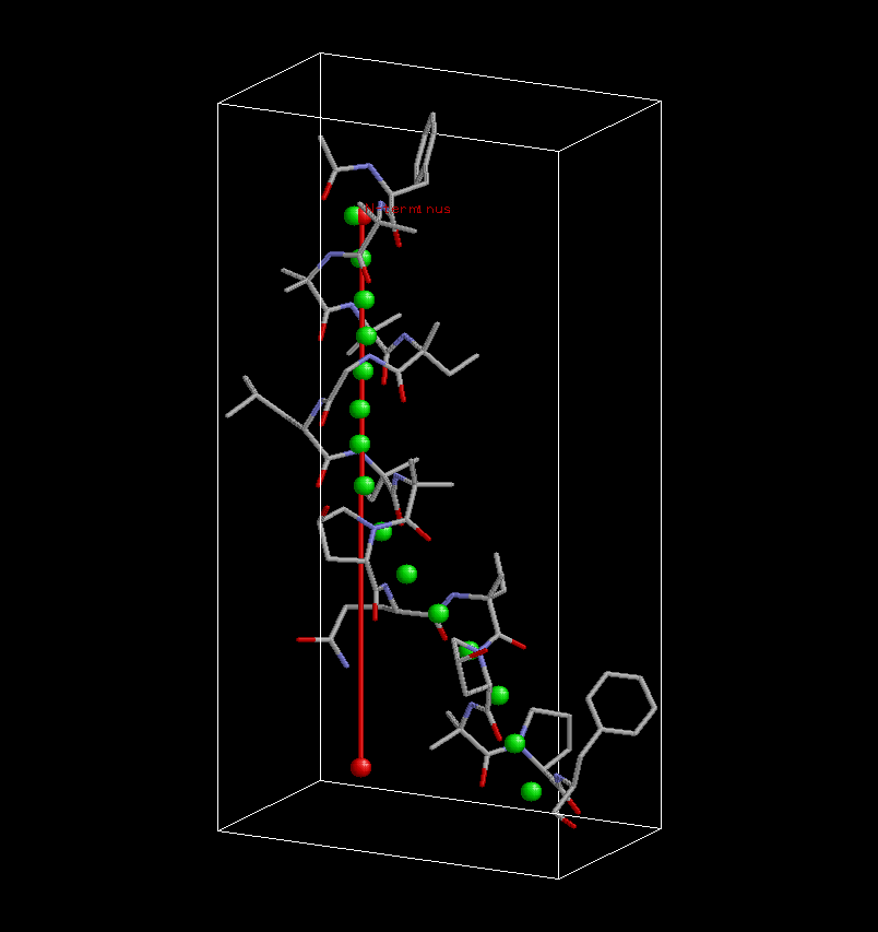
chain A
-
2D bending graph (chain A) [PDF]
-
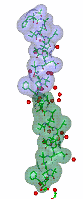
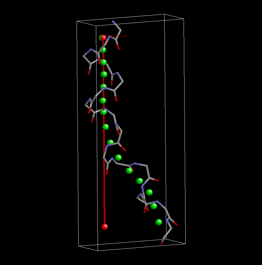
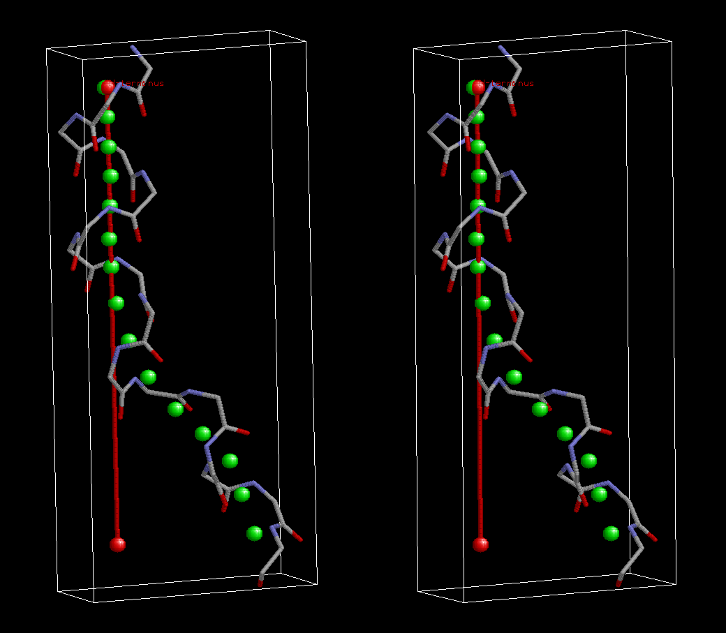
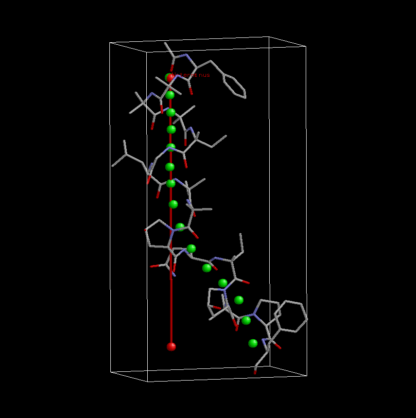
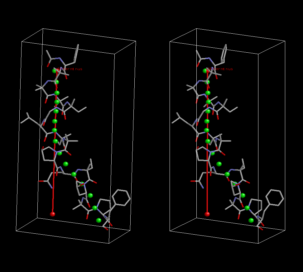
3D structure superimposed on 3D
curvilinear (green balls) and straight (red
stick) axes
-
backbone structure
-
complete structure
-
Approximate bending angle:
coming soon [read Methods
section for a critical assessment of
bending angle]
-
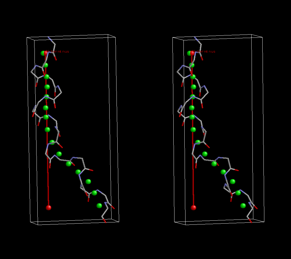
chain B
-
2D bending graph (chain A) [PDF]
-
3D structure superimposed on 3D
curvilinear (green balls) and straight (red
stick) axes
-
backbone structure
-
complete structure
-
Approximate bending angle:
coming soon [read Methods
section for a critical assessment of
bending angle]
-
Comparative bending analysis of peptaibols (1OB6 not included)
|
Reference:
Bunkoczi G, Schiell M,
Vertesy L, Sheldrick GM.
J. Peptide Sci. 2003, 9, 745-52.
Crystal structures of cephaibols.
Peptaibol
Start Page | JenaLib Home