 |
Three-Dimensional
Structures of Peptaibols |
Ampullosporin A
Sequence:
| Ac |
TRP01 |
ALA02 |
AIB03 |
AIB04 |
LEU05 |
AIB06 |
GLN07 |
AIB08 |
AIB09 |
AIB10 |
|
GLN11 |
LEU12 |
AIB13 |
GLN14 |
LEU15 |
ol |
- |
- |
- |
- |
Structural
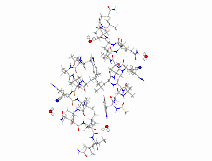
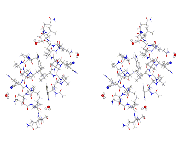
Information (X-ray structure): The asymmetric unit contains one
ampullosporin molecule, one acetonitrile molcule and two water
molecules (resolution: 0.77 Å).
 |
-
CSD
structure file in CIF format - includes two ampullosporin molecules,
two acetonitriles and four water molecules The can be requested
from the the Cambridge Structural Database by indicating the code
195231and the journal reference.
-
PDB file in PDB format generated by the authors -
includes one ampullosporin and one water molecule. Contrary to the CIF
file the PDB files includes amino acid assignments of atoms.
-
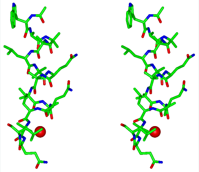
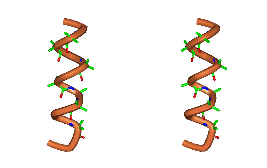
Structure
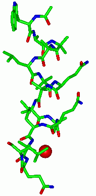
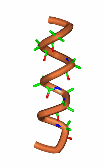
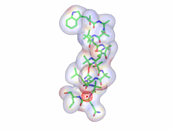
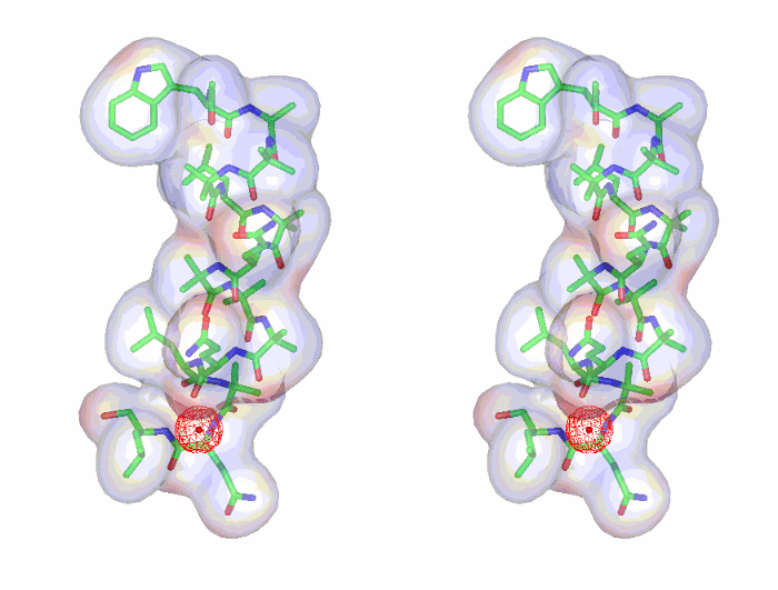
images (The thumbnail
image shows in addition to the ampullosporin structure an oxygen of one
of the two water molecules).
-
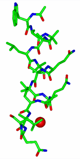
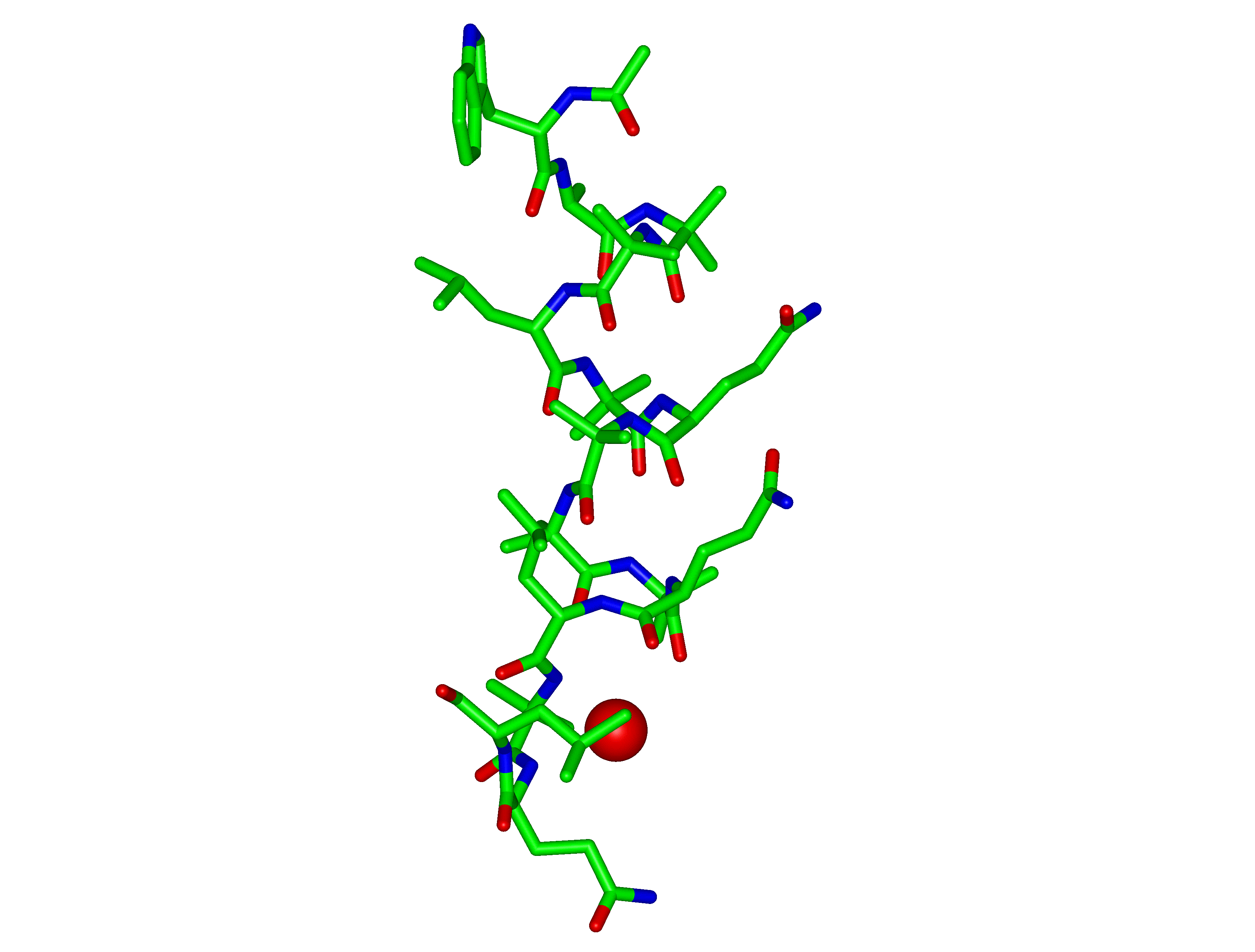
ampullosporin
A: sticks; water oxygen: spacefill (only one water molecule shown)
-
ampullosporin
A: ribbon, AIB (sticks) highlighted
-
ampullosporin
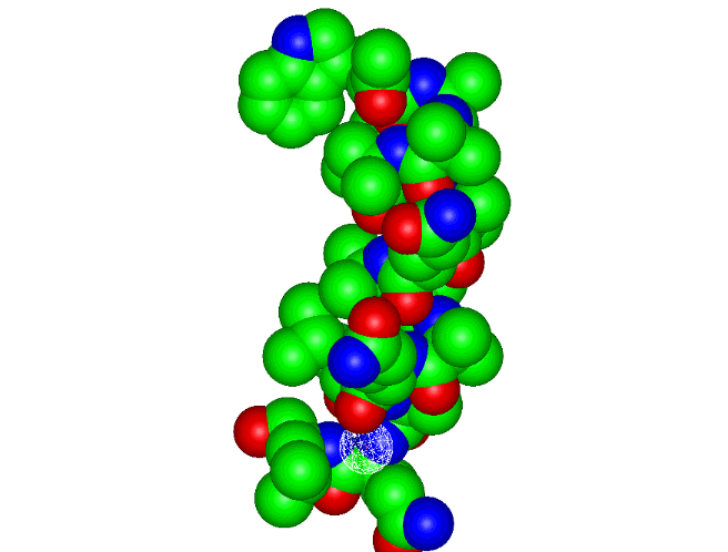
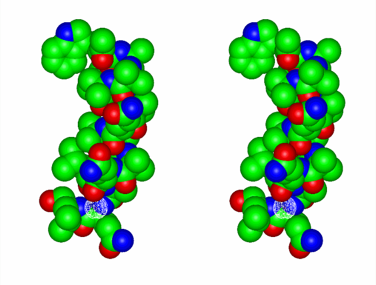
A: spacefill; water oxygen: wireframe surface (only one water molecule shown)
-
ampullosporin
A: sticks tranparent surface; water oxygen: wireframe surface (only one water molecule shown)
-
elementary unit with two ampullosporin A
molecules, 4 water molecules and two acetonitrile moelcules
-
Bending
analysis
-
Methods
-
2D
bending graph [PDF]
-
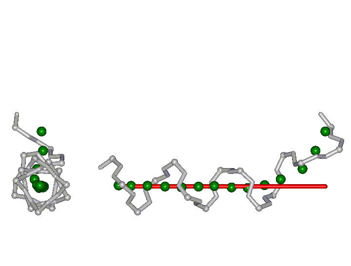
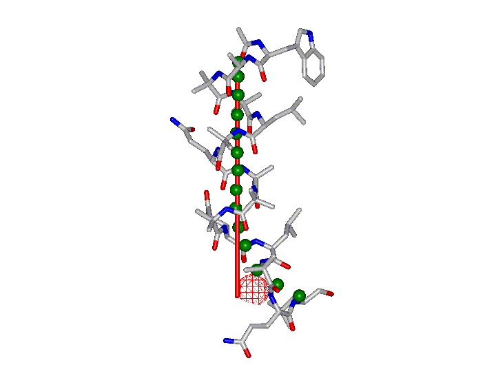
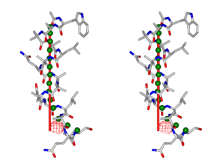
3D
structure (grey sticks, Calpa: balls) with 3D
curvilinear (green balls) and straight axes
(red stick)
-
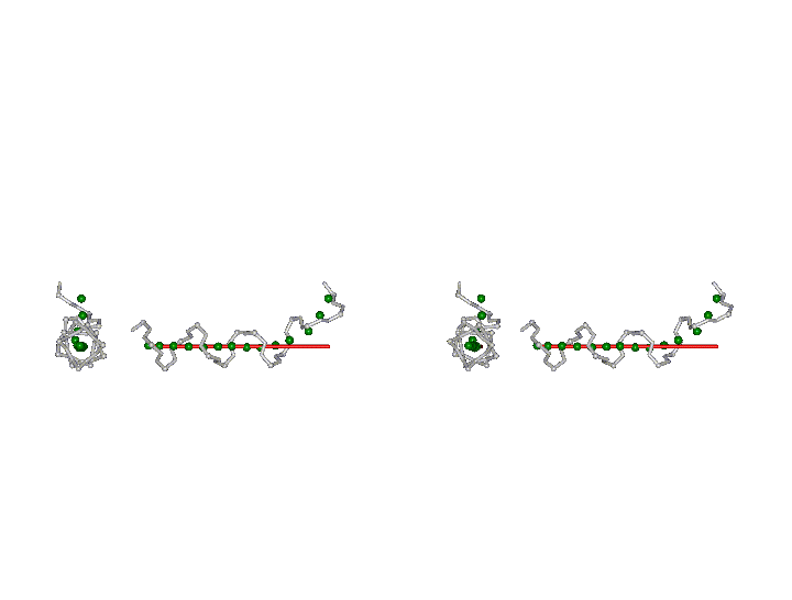
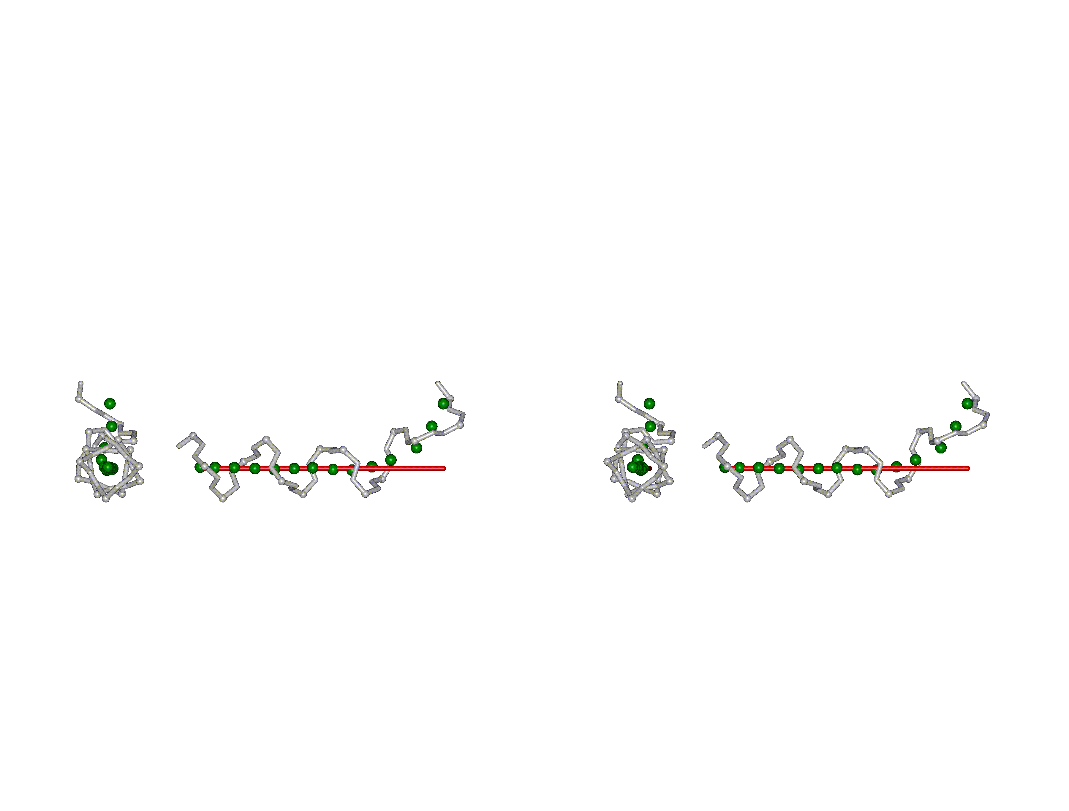
backbone structure
-
complete structure
-
Approximate bending angle: 53-64
° [read Methods section
for a critical assessment of bending angle determination and see a Table with
bending angles for all structures]
-
Comparative
bending analysis of peptaibols
|
Reference:
Kronen M, Goerls H,
Ngyuen HH, Reissmann S, Bohl M, Suehnel J, Graefe U.
J. Peptide Sci. 2003,
9, 729-744.
X-ray structure and
conformational analysis of ampullosporin A
Peptaibol Start Page | JenaLib Home