 |
Three-Dimensional Structures of Peptaibols |
[Leu1]-Zervamicin
Sequence:
| Ac |
LEU01 |
ILE02 |
GLN03 |
IVA04 |
ILE05 |
THR06 |
AIB07 |
LEU08 |
AIB09 |
HYP10 |
|
GLN11 |
AIB12 |
HYP13 |
AIB14 |
PRO15 |
PHE16 |
ol |
- |
- |
- |
Structural Information [X-ray structure; resolution ~0.93 Å]:
 |
-
CSD files (MOL2
| JNL);
CSD code: KIYPUD
-
Structure images
-
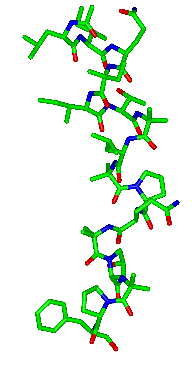
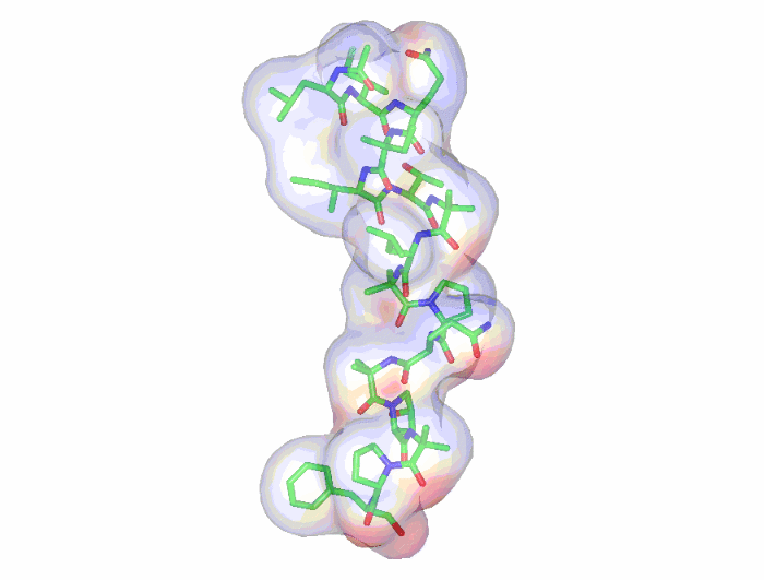
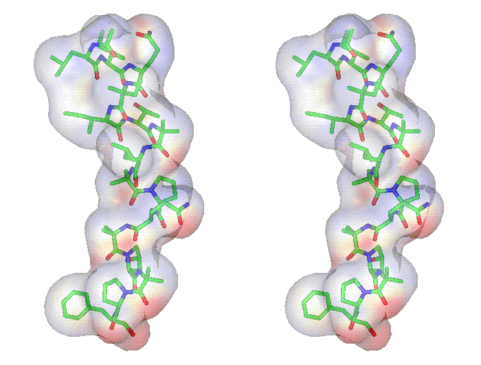
[Leu1]-zervamicin: sticks
-
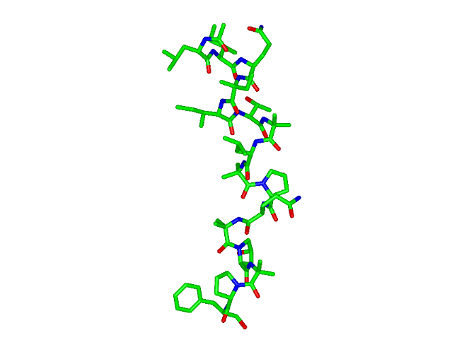
[Leu1]-zervamicin: sticks,
transparent surface
-
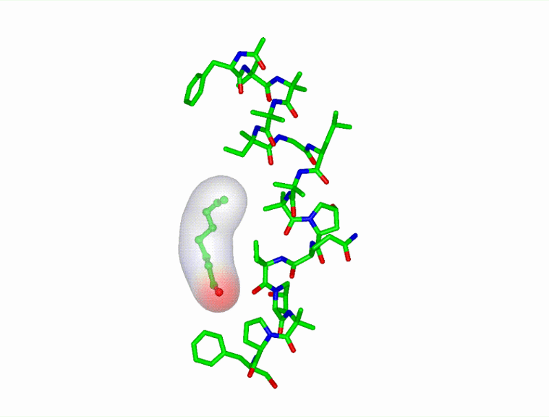
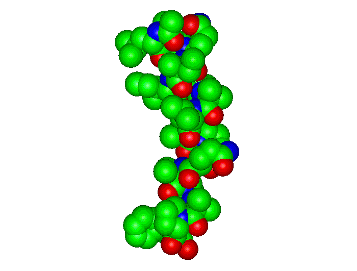
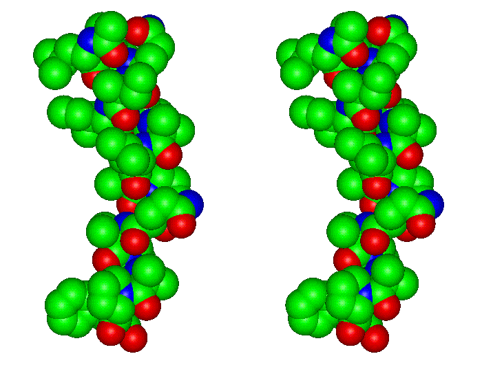
[Leu1]-zervamicin: spacefill
-
Bending analysis
-
Methods
-
2D bending graph [PDF]
-
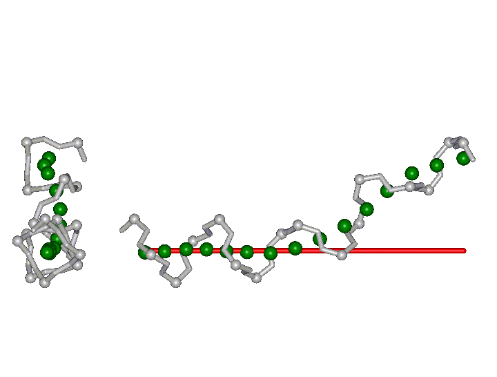
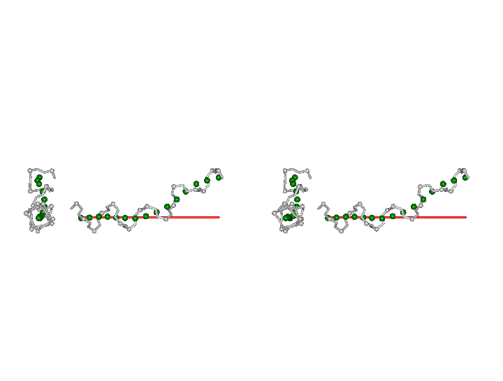
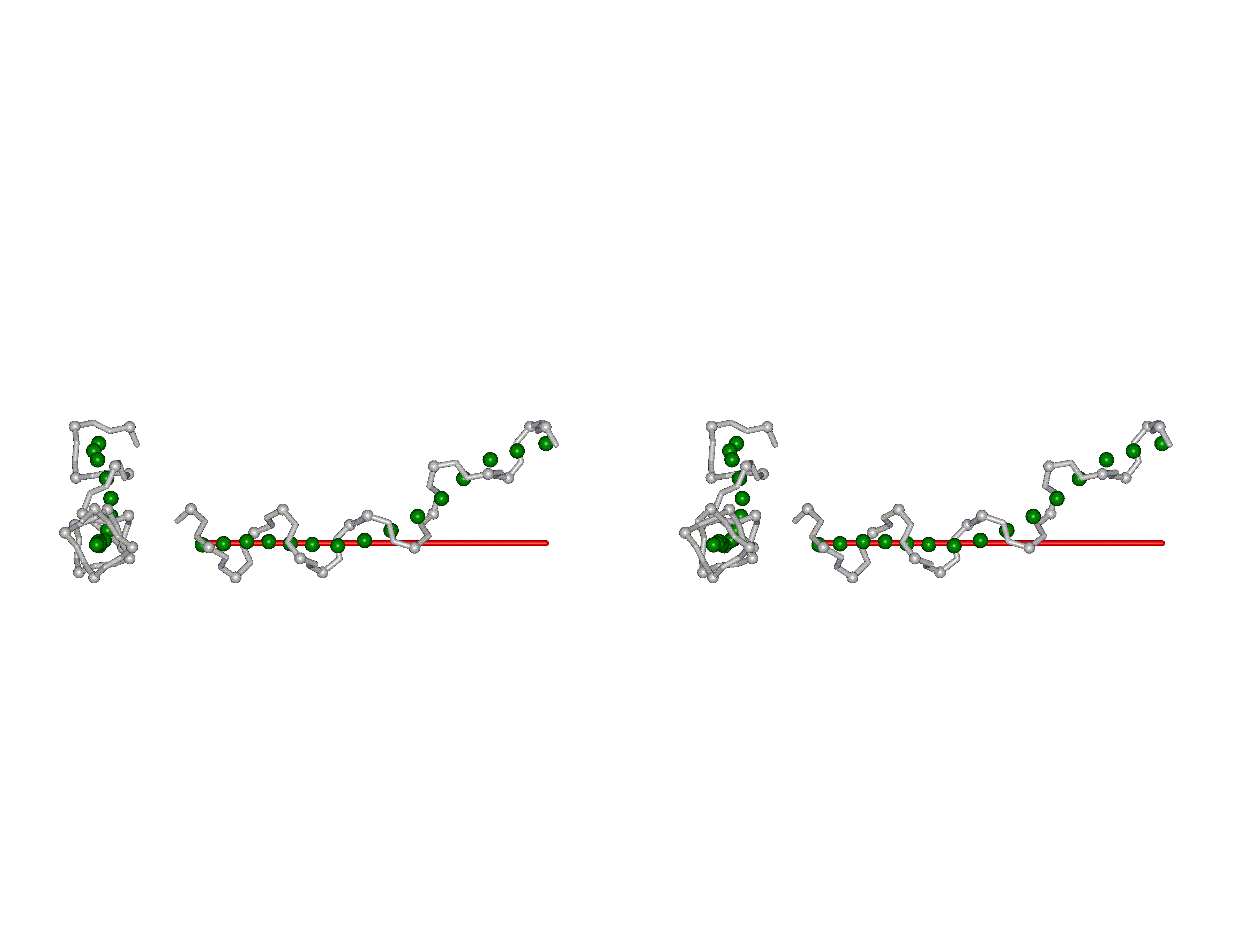
3D
structure (grey sticks, Calpha: balls) with 3D
curvilinear (green balls) and straight axes
(red stick)
-
Approximate bending angle: 31-39
° [read Methods
section for a critical assessment of bending
angle determination and see a Table
with a compilation of bending angles for all structures]
-
Comparative bending analysis of
peptaibols
|
Reference:
Karle IL, Flippen-Anderson JL, Agarwalla S, Balaram P.
Proc Natl Acad Sci U S A 1991, 88,
5307-5311.
Crystal structure of [Leu1]zervamicin, a membrane
ion-channel peptide: implications for gating mechanisms.
Peptaibol Start Page
| JenaLib Home